


Body Radiology
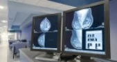
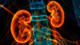
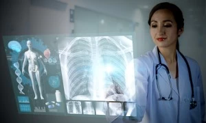
Body imaging radiologists use non-invasive diagnostic imaging to look for abnormalities inside the body and perform limited invasive procedures by guiding instruments inside the body to sample tissue. In the chest, this includes the lungs, heart and vessels. In the abdomen, this includes the liver, spleen, kidneys, pancreas, adrenal glands, vessels and intestines. In the pelvis, this includes the bladder and female organs. This has all but eliminated the need for exploratory surgeries often performed in the past.
Our Expertise
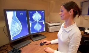
The Midwest Radiology staff has far-reaching areas of expertise, including image-guided biopsies, fluoroscopically guided procedures, women’s imaging, emergency medicine imaging, body MRI, outpatient imaging, oncologic imaging, ultrasound, thoracic radiology, abdominal radiology, sports medicine imaging, and more. Our highly trained staff has expertise in a variety of diagnostic and imaging modalities our Body Imaging division covers:
- Large tertiary care hospitals
- Emergency imaging at Level 1 trauma centers
- Oncologic imaging at cancer hospitals
- Musculoskeletal imaging at large orthopedic practices
- General outpatient imaging at several full-service outpatient imaging centers
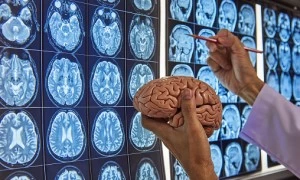
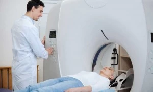
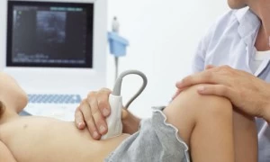
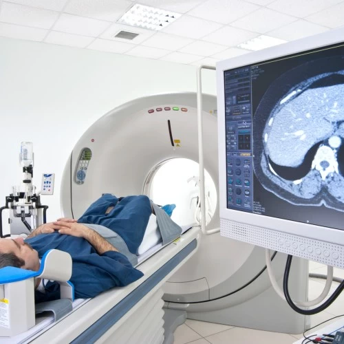
Imaging Modalities Used in Body Radiology

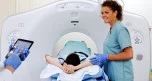
CT

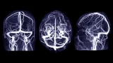
MRI

Ultrasound
Scheduling a Procedure
Midwest Radiology maintains dedicated scheduling resources for all outpatient imaging center locations. We offer same-day scheduling and accept most forms of medical insurance. Simplify your referral process, call us today!
Our Physicians
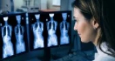
Qualified, caring, honored, and committed ─ the best of Midwest Radiology. Radiologists specializing in body imaging study organ systems of the chest, abdomen and pelvis. Body radiologists interpret images from MRI, CT, PET, ultrasound and nuclear medicine exams. Request more information about our doctors by calling 651.292.2000.