


MRI
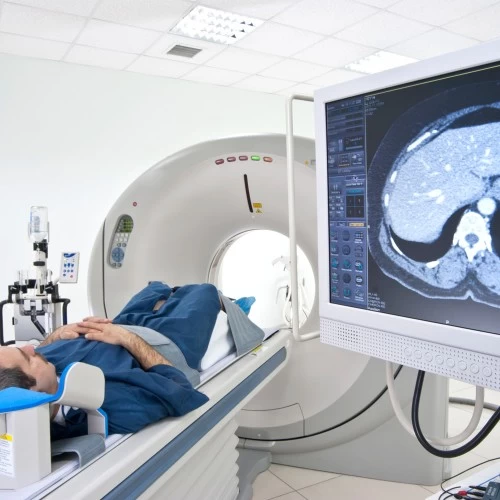
Magnetic Resonance Imaging (MRI) is a medical imaging exam that uses a strong magnetic field and radio waves to collect detailed information about the organs and soft tissues of the body. MRI does not use x-ray or radiation. MRI can assist physicians in detecting and diagnosing diseases or other abnormalities in very early stages.
What to Expect
An MRI machine temporarily realigns hydrogen atoms in the body. Radio waves make these atoms create very faint signals—and those are used to make cross-sectional images. Those images are layered on top of each other to give doctors a view of the body that they can see from different angles. MRI provides excellent anatomical detail of the soft tissues and can spot a huge range of issues, including disk abnormalities in the spine, joint problems, tumors in various organs like the kidneys and ovaries, structural problems in the heart, and brain injuries.
What is Contrast?
Your doctor may request that you receive an injection of a contrast agent called “gadolinium”. If you are having an MRI with contrast, the technologist will start an IV in your arm. Unlike contrast agents used in x-ray studies, MRI contrast agents do not contain iodine and rarely cause allergic reactions or other problems. This contrast enhances the image quality, allowing the radiologist to be more accurate and confident in their diagnosis. When the imaging exam is complete, the contrast material is either absorbed by the body or eliminated through urine.
MRI Procedures

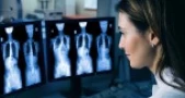
Brain MRI

MRI Lumbar Spine

MRI of Joint Lower Extremity

MRI Cervical Spine

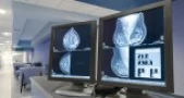
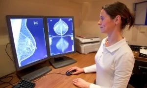
Breast MRI

MR Angiography (MRA)

MRI Joint Upper Extremity

MRI Arthrogram

MRCP

MR Enterography
Wide Open Spaces
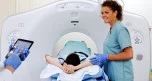
Wide Bore MRI provides the most comfortable advanced imaging experience.
Wide Bore Magnetic Resonance Imaging, or wide bore MRI, is a very patient-friendly scan that is quieter and more comfortable for patients than a standard MRI. Wide bore MRI gathers information about your body using a magnet, radio waves, and a computer to create detailed internal images of the body and transmits them for your doctor to see.
Claustrophobic and obese patients as well as those feeling anxious or concerned are often able to comfortably undergo wide bore MRI examinations. It features a large and comfortable bed and opening, reduced noise and allows patients to relax during the exam. In addition to enhanced comfort, this technology features high-field technology to provide outstanding image quality.
Scheduling a Procedure
Midwest Radiology maintains dedicated scheduling resources for all outpatient imaging center locations. We offer same-day scheduling and accept most forms of medical insurance. Call us today!
Locations
Our outpatient imaging services are provided through our network of imaging centers that are conveniently located throughout the Twin Cities area. We offer top-quality imaging services that generate high-resolution imagery using state-of-the-art medical imaging equipment.
Store Locator is loading from Storemapper...
Our Physicians
Qualified, caring, honored, and committed ─ the best of Midwest Radiology. Our radiologists are board certified by the American Board of Radiology and have extensive training and expertise in medical imaging. They are dedicated to providing the highest level of quality imaging services to all patients and healthcare providers.